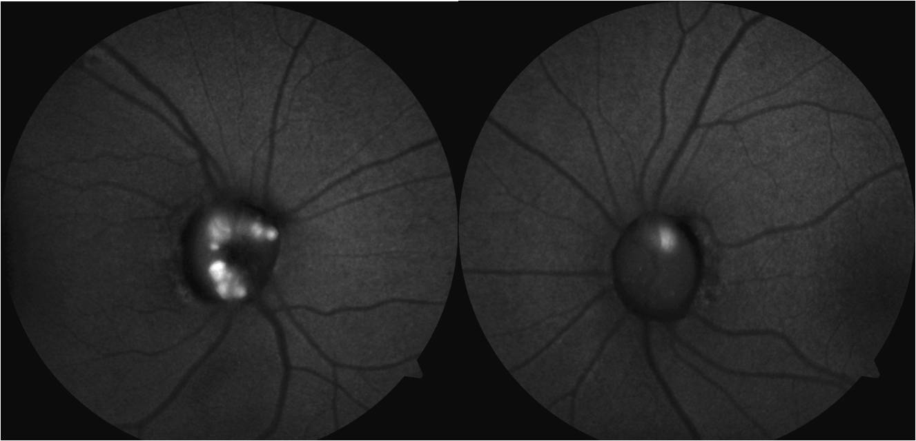
These are hyaline bodies which can calcify at the optic nerve head, and are seen during slit lamp fundus biomicroscopy.
A particular characteristic is autofluorescence of the drusen – seen above – using the correct filters, there is no need for any fluorescent molecules to help identify them.
Some patients with optic disc drusen may have transient visual obscurations. Rarely, arcuate visual field defects and increased blind spot in visual field testing are seen, as well as non-arteritic ischemic optic neuropathy and retinal vein occlusions.